Press release
Thursday, March 11, 2021
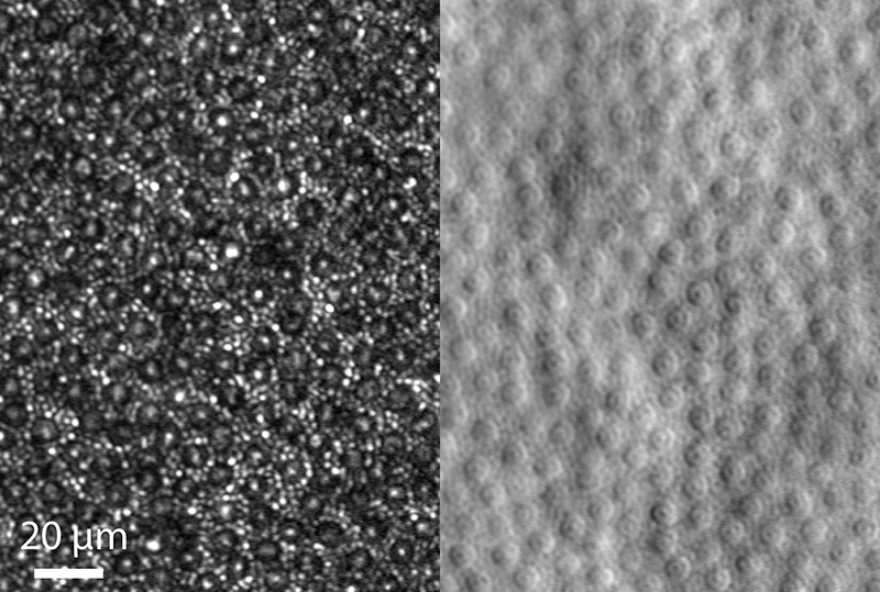
By removing the strange light, the scientists improved the resolution by 33%.
A team led by scientists at the National Eye Institute (NEI) has non-invasively visualized cells that detect light in the back of the eye, known as photoreceptors, in more detail than ever. Published in Optica, the researchers report how they improved image resolution by a third by selectively blocking the light used to imagine the eye. NEI is part of the National Institutes of Health.
The achievement is the latest in an evolving strategy to control cellular changes in retinal tissue that, in turn, will help identify new ways to treat and prevent vision loss from diseases such as macular degeneration related to age, one of the main causes of blindness over the age of 65.
“Better image resolution will allow better monitoring of degenerative changes that occur in retinal tissue. The goal of our research is to discern changes related to disease at the cellular level over time, possibly allowing the detection of the disease much earlier, ”said the study’s lead researcher, Johnny Tam, Ph.D. Unit at NEI.
Prior detection would allow patients to be treated earlier, long before they lost their vision. In addition, detecting cellular changes would allow doctors to more quickly determine if a new therapy works.
The two types of photoreceptors, the cones, which allow color vision, and the bars, which allow vision in low light, vary in size and density in the retina. Cone photoreceptors, although larger than bars, are more difficult to visualize when better packaged because they are located in the fovea, the region of the retina responsible for the highest level of visual acuity and color discrimination. . The whole landscape of cones and rods is known as the photoreceptor mosaic.
Advanced imaging systems are widely used to observe retinal tissue and are essential tools for diagnosing and studying retinal diseases. But even with retinal imaging of adaptive optics, a technique that compensates for light distortions using deformable mirrors and computer-driven algorithms, there are still some areas of the photoreceptor mosaic that are difficult to image, according to the first author of the document, Rongwen Lu. , Ph.D., postdoctoral fellow in the Clinical and Translational Imaging Unit of NEI.
“Sometimes the bars are hard to imagine because they’re so small,” Lu said. “By removing some of the light from the system, it’s actually easier to see the bars. Therefore, in this case, less is more ”.
In this latest report, Tam’s team at NEI, with the help of researchers from Stanford University, Palo Alto, California, tried to further drive the resolution of the adaptive optic retina image by strategically blocking part of the light to imagine the retina.
By blocking the light that illuminates the eye in the middle of the beam, to create a ring of light (instead of a disk), the NEI-led equipment improved the transverse resolution (through the mosaic). But this happened at the expense of axial resolution (mosaic depth). To compensate, Tam’s team blocked the light returning from the eye through a very small hole, called a sub-aerated disk, which recovers the axial resolution that would have been lost with just the light ring.
The combination of ring lighting with the sub-Airy disc image results in the best of both worlds, Tam said. The tight technique provides a 33% increase in resolution, making it much easier to see the bars as well as the subcellular details inside the cones.
His technique also improved the visualization of the photoreceptor mosaic with another technique called detection of non-confocal fractions, which is another type of microscopy that provides a complementary view of the photoreceptor mosaic.
The work was supported in part by grants NEI U01 EY025477 and R01 EY025231, and by the NEI Intramural Research Program, which is part of the National Institutes of Health.
NEI is leading federal government research on the visual system and eye diseases. NEI supports basic and clinical science programs to develop vision-saving treatments and meet the special needs of people with vision loss. For more information, visit https://www.nei.nih.gov.
Regarding the National Institutes of Health (NIH):
NIH, the country’s medical research agency, includes 27 institutes and centers and is a component of the U.S. Department of Health and Human Services. NIH is the leading federal agency that conducts and supports basic, clinical, and translational medical research and investigates the causes, treatments, and cures for common and rare diseases. For more information about NIH and its programs, visit www.nih.gov.
NIH … Turning discovery into health®
References:
Lu R, Aguilera N, Liu T, Liu J, Giannini JP, Li J, Bower AJ, Dubra A, Tam J. “Images of adaptive in vivo sub-diffraction optics of photoreceptors in the human eye with illumination of annular pupil and sub-airy detection ”, published on March 11, 2021, Optica. https://doi.org/10.1364/OPTICA.414206